
- Medical Specialities
-
- Ahmedabad
- Vadodara
- Anand
-
Call Us
For booking appointments and health check up queries
+91 99 09 02 1667
For Medical Emergency
+91 79 6619 0200
For Information and Inquires
+91 79 6619 0201
For International Patient Inquiry
+91 99090 21667

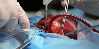
Cardiac Surgery
Medical Specialities / Cardiac Surgery
FAQ
Carotid artery disease
Carotid artery disease occurs when the major arteries in your neck become narrowed or blocked. These arteries, called the carotid arteries, supply your brain with blood. Your carotid arteries extend from your aorta in your chest to the brain inside your skull.
Your arteries are normally smooth and unobstructed on the inside, but as you age, a substance called plaque can build up in the walls of your arteries. Plaque is made up of cholesterol, calcium, and fibrous tissue. As more plaque builds up, your arteries narrow and stiffen. This process is called atherosclerosis, or hardening of the arteries.
Carotid artery disease may not cause symptoms in its early stages. Unfortunately, the first sign of carotid artery disease could be a stroke or a transient ischemic attack.
Lifestyle changes to lower your risk include:
- Stop smoking
- Control diabetes
- Control blood pressure
- Be physically active
- Eat a low-saturated-fat, low-cholesterol diet

Your treatment will depend on the severity of your condition, and whether or not you are having symptoms from the carotid artery disease, as well as your general health. As a first step, your physician may recommend medications and the lifestyle changes.
Carotid Artery Stenting is a non-surgical procedure that can be used to dilate (widen) narrowed or blocked carotid arteries. A tiny coil (stent) is expanded inside the blocked artery and is left in place to keep the artery open. Carotid artery stent procedures are performed with special devices (embolic protection devices) to prevent plaque from entering the circulation of the brain during the stent procedure.
Structural heart disease most often refers to cardiac defects which are congenital in nature (birth defects), but may also include abnormalities of the valves and vessels of the heart wall that develop with wear and tear on the heart, or through other disease processes. The three most common congenital heart diseases are atrial septal defect (ASD), patent foramen ovale (PFO), and coarctation of the aorta.
An ASD is a hole in the wall (septum) which separates the top two chambers of the heart. A PFO is similar to an ASD; it is a flap-like hole in the wall that separates the upper two chambers of the heart, a coarctation of the aorta is a narrowing of the vessel, in the upper chest that carries the blood from the heart, to every other part of the body to supply oxygen and other nutrients.
What are Symptoms of Structural Heart Disease?
-
In PFOs
- Transient Ischemic Attack (TIA or mini-stroke)
- Migraine headaches
- Low oxygen levels in rare patients.
- “Bends” in divers
-
In ASDs
- Heart palpitations
- Exercise intolerance
- Stroke
There are treatments in the cath lab treatment for ASDs, PFOs and coarctations. For both ASDs and PFOs there is a catheter based procedure that utilized specific closure devices. Stenting is used to treat patients coarctations.
Valvular heart disease
Heart valves are flaps, or leaflets, of tissue that ensure that blood entering or leaving the heart moves in the proper direction with no backflow. The heart has a total of four valves, the mitral, triscupid, aortic and pulmonary valve. Valvular disease can affect any of these four valves, and can interfere with the normal flow of blood through the heart Valvular stenosis, or a narrowing of the valve requires the heart to pump harder, which can strain the heart and reduce blood flow to the body. The tissues forming the valve leaflets become stiffer, narrowing the valve opening and reducing the amount of blood that can flow through it. If the narrowing is mild, the overall functioning of the heart may not be reduced. However, the valve can become so narrow (stenotic) that heart function is reduced, and the rest of the body may not receive adequate blood flow. Valvular regurgitation (incompetent, insufficient, or leaky) valve does not close completely, letting blood move backward through the valve. This backward flow is referred to as “regurgitant flow.”
There are many types of valve disease .Valve disease can be congenital (present at birth) or may be acquired later in life. Acquired valve disease includes problems that develop with valves that were once normal. These may involve changes in the structure of your valve or infection, such as rheumatic fever which causes a the heart valve leaflets to become inflamed, and may cause the leaflets to stick together and become scarred, rigid, thickened and shortened. Other causes of valve disease include: coronary artery disease, heart attacks, cardiomyopathy (heart muscle disease), syphilis, hypertension, aortic aneurysms, connective tissue diseases, and less commonly, tumors some types of drugs and radiation.
If the valve disease is more serious, the symptoms can include:
- Breathlessness during exertion
- Waking up at night short of breath
- Palpitations (irregular, fluttery heartbeat)
- Angina (chest pain) because the blood vessels supplying the heart muscle are not getting enough blood
- Swelling of the ankles or feet
- Tiredness
- Dizziness or fainting
Treatment depends on what is wrong with the valve and how serious the problem is. If the disease is minimal, medical therapy may be prescribed to relieve the symptoms.
Balloon Valvuloplasty is a procedure in which the narrowed heart valve is stretched open. A small incision is made in the skin and a catheter is inserted into an artery in the leg, where a balloon-tipped catheter is advanced into the heart and across the narrowed valve. When in place, the balloon is expanded to open the valve, resulting in improved blood flow across the diseased valve.
Percutaneous mitral valve repair (MVR) is a new treatment being investigated to determine the effectiveness of a percutaneous approach for treating mitral valve regurgitation. The procedure is performed in the cardiac catheterization laboratory with the aid of echocardiography. During this procedure, a very small, specially made metal clip device is delivered through a catheter inserted into the femoral vein (in the groin) and advanced to the heart. Guided by echocardiography, the cardiologist attaches the clip to the flaps of the mitral valve. Placement of the clip is adjusted until optimal improvement in blood flow and pressures through the valve are observed. Then, the clip is released, and the catheter is withdrawn. The clip holds the valve flaps in position, which limits the leakage.
Our Cardiothoracic Surgery department delivers outstanding level of surgical care to patients suffering from heart diseases. The department is a leader in performing complicated heart surgeries, such as the Coronary Artery Bypass Grafting (CABG, or Heart Bypass Surgery), Valvuloplasty and other complex procedures. It has an exceptional record of high safety and success rate for complex heart surgeries. It is led by renowned surgeons and highly skilled staff, and supported by a state-of-the-art facility with ultra modern equipment. The department provides around the clock services to patients requiring surgical care.
Our Cardiothoracic Department is equipped with the latest equipment and facilities
Operation Theatres
- We have four dedicated operation theatres, for the Cardio-Thoracic department. Our operating theatres are fully equipped and prepared for all types of heart surgeries, including Beating Heart Surgery (Bypass Off Pump), Open Heart Surgery (Bypass On Pump), Valvular and Cogenital Surgery, Pulmonary and other complex procedures.
- Filtration of air in the operating theatres is critical for preventing infections. Our theatres have Micro-V and Hepa filters to ensure sanitary air supply during the procedures. The air is changed every 2 minutes and a positive pressure is maintained to prevent outside air from coming in.
- All materials of our operating theatres, including floor and wall materials, are non-porous to prevent growth of any bacteria or infectious materials. These washable materials are kept sterile by top quality of sterilization and disinfection processes. The operating rooms are built to allow every nook and corner to be kept easily sterile.
- Our operating theatres are furnished with the latest equipments
We provide exceptional high quality post operative care in our well-equipped and dedicated Intensive Cardiac Care Units (ICUs) for the Cardiothoracic. We have one of the lowest incidences of post operative infections in the nation. We maintain a 1:1 nurse to patient ratio to ensure positive outcomes and caring service.
CABG (Coronary artery bypass graft)
Operation for CABG is designed to bypass the blockages in the coronary arteries in order to restore normal or near normal blood flow to the entire heart during rest and exercise. Though an occasional patient needs only one bypass graft, most people who are candidates for CABG have blockages in most of their coronary arteries and need between three and five bypass grafts.
In this surgery, a healthy blood vessel is taken (artery or vein from your body) usually from the leg, arm, chest or abdomen, and connected to the other arteries (usually the aorta) in the heart. This enables blood flow to “bypass,” or go around, the diseased or blocked portion of the coronary artery, creating a new path for blood flow to the heart.
To begin the CABG operation,an incision is made down the front of the chest, usually dividing the breastbone or sternum. This incision is called a median sternotomy, and it enables the surgeon to safely operate on all parts of the heart. During the surgery, the patient typically will be connected to a heart-lung machine (sometimes called “the pump”), which is a machine that temporarily takes over the function of the heart and lungs during surgery to maintain blood circulation and oxygen flow through the body. The heart-lung machine allows heart to be stopped in order to carefully and accurately sew the bypass grafts to the heart. After the surgery is completed, the patient will be taken off the pump, and his/her heart and lungs will resume their function.
After CABG, the patients spend 3 nights in an intensive care unit, followed by another 5-6 days in the hospital.x
CABG can also be performed without the use of the heart lung machine (“off pump”) by sewing the coronary bypass grafts to the coronary arteries with the heart beating. This technique requires the use of instruments that “stabilize” or hold the area of the coronary artery being bypassed still so that the surgeon can carefully and accurately sew the graft to it.
Mitral valve replacement
Severe valve damage means the valve must be replaced and most often involves the mitral valve. It is also used to treat any valve disease that is life-threatening. Some patients may need more than one valve repaired or replaced.
Two kinds of valves can be used for replacement:
- Mechanical valves are usually made from materials such as plastic, carbon, or metal. Mechanical valves are strong, and they last a long time. Because blood tends to stick to mechanical valves and create blood clots, patients with these valves will need to take blood thinning medicines (anticoagulants) for the rest of their lives
- Biprosthetic valves are made from animal tissue (called a xenograft) or taken from the human tissue of a donated heart (called an allograft or homograft). Sometimes, a patient's own tissue can be used for valve replacement (called an autograft). Patients with biological valves usually do not need to take blood-thinning medicines.However, the bioprosthetic valves may only last 10 to 15 years.[6] The choice of which valve type to use depends upon the patient's age, medical condition, preferences with medication, and lifestyle
During valve repair or replacement surgery, the breastbone is divided, the heart is stopped, and blood is sent through a heart-lung machine. Because the heart or the aorta must be opened, heart valve surgery is open heart surgery.
Aneurysms
An aneurysm is a balloon-like bulge in an artery. Aneurysms can form in arteries of all sizes and occurs when the pressure of blood passing through part of a weakened artery forces the vessel to bulge outward. All aneurysms are not life threatening unless, the bulging stretches the artery too far, this vessel may burst, causing a person to bleed to death. An aneurysm that bleeds into the brain can lead to stroke or death.
Aneurysms occur in parts of the large vessel that carries blood from the heart to other parts of the body (the aorta). Aortic aneurysms can occur in the area below the stomach (abdominal aneurysms) or in the chest (thoracic aneurysms). An abdominal aortic aneurysm (AAA) is usually located below the kidneys. It can also occur in the heart's main pumping chamber (the left ventricle).
Open-chest surgery to treat a thoracic aortic aneurysm is to replace the weakened portion of the aorta with a fabric tube, called a graft. The surgeons will have to open either the stomach area (for abdominal aneurysms) or the chest (for thoracic aneurysms). When the operation begins, the surgeon will make a cut (called an incision) either in the abdomen or the chest depending on the location of the aneurysm.Clamps would be placed on the artery above and below the aneurysm to stop blood flow to that part of the aorta.
The bulging section of the aorta is cut out (excised). The surgeon will then replace the missing piece with a fabric tube called a graft. The clamps are then removed slowly to allow blood to flow through the vessel again.
Sometimes surgeons do not cut out the bulging section of the aorta. In these cases, the operation is the same except that surgeons place the fabric graft inside the vessel, like a lining, to decrease the pressure on the wall of the artery. This procedure is called endo-aneurysmorrhaphy. If the aneurysm is located in the ascending aorta just above the heart, the heart-lung machine will be used.
Open surgery to repair an abdominal aortic aneurysm is sometimes done as an emergency procedure, you may have an abdominal aortic aneurysm that is not causing any symptoms or problems and had been found after an Ultrasound or CT done for another reason. There is a risk that this aneurysm may suddenly break open (rupture) if you do not have surgery to repair it.
Aortic valve replacement
Surgery to replace an aortic valve is done for aortic valve stenosis and aortic valve regurgitation. During this surgery, the damaged valve is removed and replaced with an artificial valve. The valve replacement is typically an open-heart surgery.
A minimally invasive surgery or a catheter procedure to replace the aortic valve may be an option for some people.
You will recover in the hospital until you are healthy enough to go home. Depending on your overall health, you will likely go home a few days after surgery.
Surgery will involve a long recovery over several weeks. You will probably need to take 4 to 12 weeks off from work. It depends on the type of work you do and how you feel. In some cases, full recovery may take several months.
Aortic valve regurgitation
If your chronic regurgitation is getting worse and you have symptoms, you will likely have surgery. You might have surgery before you get symptoms, especially if your regurgitation is getting worse. If you have acute regurgitation, surgery will likely be done right away.
Valve replacement is recommended based on many things including how severe the stenosis is, whether you have symptoms, and how well your heart is pumping blood. It is typically recommended when a person has severe stenosis.
Valve replacement surgery can fix aortic valve regurgitation. It helps relieve symptoms and prevent heart failure. And it helps people live longer.
Valve replacement surgery is an effective treatment for people who have severe aortic valve stenosis.2 If you don't have surgery after your stenosis is severe, you may die suddenly or develop heart failure. Surgery can relieve symptoms, improve your quality of life, and help you have a more normal life span.
Valve replacement surgery has a high rate of success and a low risk of causing other problems if you are otherwise healthy. Although most people have successful outcomes, there is a risk of death and serious problems during surgery. Valve replacement surgery is high-risk for people who have a failing left ventricle and who have had a heart attack. About 5 or less out of 100 people who have valve surgery die.3
If you have severe aortic valve regurgitation or stenosis, the risks of not replacing the valve may be greater than the risks of surgery, unless you have other health problems that make surgery too dangerous.
Tricuspid valve repair
Tricuspid valve repair and tricuspid valve replacement are procedures that treat diseases affecting the tricuspid valve.
The tricuspid valve is one of four valves that regulate blood flow through the heart. These valves keep blood flowing in the right direction through the heart.
The tricuspid valve separates one of the heart's two upper and lower chambers (atria and ventricles). With each heartbeat, the atria fill with blood from the body and lungs, and the ventricles contract to pump blood to the lungs and the rest of the body.
As the atria fill to capacity, the tricuspid valve opens to allow blood to flow from the right atrium into the right ventricle. As the ventricles contract, the tricuspid valve shuts tightly to prevent blood from flowing back into the right atrium.
When the tricuspid valve isn't working properly, it can interfere with the proper direction of blood flow and force the heart to work harder to supply the necessary blood to the lungs and the rest of your body.
Tricuspid valve disease is often caused by a heart defect present at birth (congenital heart disease) and may require immediate medical attention in infants.
For others, tricuspid valve disease may not cause any signs or symptoms for many years, if at all. Some people may experience pulsations in the neck, abdominal or chest pain, shortness of breath with activity, fatigue, irregular heartbeat (arrhythmia), heart failure, or sudden cardiac death.
Tricuspid valve repair or tricuspid valve replacement can treat tricuspid valve disease and help restore normal blood flow, reduce symptoms, improve survival in some people and help preserve the function of your heart muscle.
Tricuspid valve disease treatment depends on the severity of your condition, whether or not you're experiencing signs and symptoms, and if your condition is getting worse.
For some people with tricuspid valve disease without any symptoms, regular monitoring under a doctor's supervision may be all that's needed.
Types of tricuspid valve disease that may require treatment with tricuspid valve repair or replacement include:
Tricuspid valve regurgitation occurs when the tricuspid valve doesn't close properly and allows blood to flow back into the right atrium when the right ventricle contracts rather than in the normal, one-way direction from the atrium to the ventricle.
The most common cause of tricuspid regurgitation is enlargement of the right ventricle due to other heart or lung disorders, such as heart failure, pulmonary hypertension or emphysema. These conditions make it harder for the blood to flow out of the right ventricle and to the lungs. To compensate for the increased resistance, the tricuspid valve dilates or stretches and can't close completely.
Tricuspid valve regurgitation may also be caused by congenital heart disease (including Ebstein anomaly), injury, infection of the heart valves (infective endocarditis) associated with the use of illicit drugs or bacterial infection (rheumatic fever).
Tricuspid valve stenosis causes the tricuspid valve to become narrowed or obstructed, which makes it more difficult for blood to flow from the right atrium to the right ventricle. Tricuspid valve stenosis may also be accompanied by tricuspid regurgitation or backflow.
Tricuspid valve stenosis may be caused by congenital heart disease, thickening of the valve's closure flaps (leaflets), carcinoid heart disease or as a result of rheumatic fever.
Tricuspid atresia is a type of congenital heart disease that occurs when a baby is born without a tricuspid valve or opening to allow blood to flow from the right atrium to the right ventricle. As a result, the right ventricle is not fully developed and surgery is often needed to increase blood flow to the lungs.
Severe tricuspid valve disease caused by congenital heart disease may require immediate and ongoing medical or surgical care in children. But other types of tricuspid valve disease may be acquired or progress and require medical intervention in adulthood.
Most tricuspid valve conditions are mechanical problems that cannot be adequately treated with medication alone and will eventually require surgery to reduce symptoms and the risk of complications, such as heart failure.
The decision to repair or replace a damaged tricuspid valve depends on many factors, including:
- The severity of your tricuspid valve disease
- Your age and overall health
- Whether you need heart surgery to correct another heart problem in addition to tricuspid valve disease, such as mitral or aortic valve repair or replacement or coronary artery bypass surgery to treat coronary artery disease, so both conditions can be treated at once
In general, heart valve repair is usually the first choice because it's associated with a lower risk of infection, preserves and optimizes heart function, and may reduce the potential need for long-term use of blood-thinning medications compared with heart valve replacement.
But not all valves can be repaired, and some repaired valves may eventually require replacement. In addition, heart valve repair surgery is often harder to perform successfully than valve replacement surgery.
Tricuspid valve repair and tricuspid valve replacement risks vary depending on your health, the type of procedure and the expertise of the health care team. To minimize potential risk, tricuspid valve surgery should generally be performed at a medical center with staff experienced in these procedures and that performs high volumes of tricuspid valve operations.
Risks associated with tricuspid valve repair and tricuspid valve replacement surgery may include:
- Bleeding
- Blood clots
- Valve dysfunction in replacement valves (valve prostheses)
- Heart rhythm problems
- Infection
- Stroke
- Death
Overall, the long-term survival rates after tricuspid valve repair and tricuspid valve replacement are similar.
Bentall’s surgery
The aorta is the largest artery that stems from the heart and branches into smaller arteries to distribute oxygenated blood to all parts of the body. It can be divided into the ascending aorta (part that rises from the heart), aortic arch (part that bends over the heart), descending thoracic aorta (part that moves down the chest region) and abdominal aorta (part that begins at the diaphragm).
The Bentall procedure is a surgery performed to correct defects of the aorta. The procedure involves the replacement of the aortic root (base of the aorta) and valve (three flaps that ensure the one-way flow of blood from the heart to the aorta), and re-implantation of the coronary arteries (that branch out from the ascending aorta). The current and most common type of surgery is called the button Bentall surgery.
- The Bentall surgery is indicated for the following conditions of the aorta
- Aortic regurgitation (leaking of the aortic valve)
- Marfan’s syndrome (genetic disease that causes aortic wall weakness)
- Aortic dissection (separation of the layers of the aortic wall)
- Aortic aneurysm (enlargement of the aorta)
The Bentall surgery is performed under general anaesthesia. During the surgery, an incision is made along the middle of the chest to expose the heart. The heart’s activity is temporarily stopped so your surgeon can perform the operation, and blood flow is redirected to a bypass machine.
The diseased part of your aorta is excised and the valve is inspected. The diseased aortic valves are removed. The coronary arteries, along with a large button of the surrounding wall of the aorta are dissected. An artificial graft that has a mechanical or bioprosthetic valve is sutured to the ends of the aorta. Two holes are created in the graft, into which the coronary artery buttons are sewn. The valve replacement may be tested by your surgeon by streaming some blood into the area and checking for any kind of leakage. Once the repair is complete, the incision is sutured and bandaged. The entire procedure is normally completed in less than 5 hours.
After the surgery, you will be taken to the post anaesthesia care unit and connected to a ventilator where your vitals will be monitored. Before discharge, your doctor will give you a set of instructions to be followed such as:
- Increase your activity progressively as tolerable
- Avoid any vigorous activity for up to 12 weeks after the surgery
- Do not lift any kind of heavy weights for the first few weeks after the surgery
Please inform your doctor if you experience the following:
- Chills
- High fever
- Drainage from the incision
- Incisional redness
- Increased incisional tenderness
As with all surgical procedures, the Bentall procedure may be associated with certain complications. Some of these complications include:
- Arrhythmias (irregular heartbeat)
- Pneumonia (lung infection)
- Septic shock (severe infection and inflammation)
- Bleeding
- Wound infections
- Disturbances in conduction and pace
- Neurological disorder
- Hepatic and renal insufficiency (failure of liver and kidney, respectively)
Your doctor will thoroughly consider your condition, and the benefits and possible risks of the Bentall procedure before proceeding with it.
Transesophageal echocardiography
Transesophageal echocardiography (TEE) is a test that produces pictures of your heart. TEE uses high-frequency sound waves (ultrasound) to make detailed pictures of your heart and the arteries that lead to and from it. Unlike a standard echocardiogram, the echo transducer that produces the sound waves for TEE is attached to a thin tube that passes through your mouth, down your throat and into your esophagus. Because the esophagus is so close to the upper chambers of the heart, very clear images of those heart structures and valves can be obtained.
- TEE is a test that uses sound waves to make pictures of your heart’s muscle and chambers, valves and outer lining (pericardium), as well as the blood vessels that connect to your heart
- Doctors often use TEE when they need more detail than a standard echocardiogram can give them
- The sound waves sent to your heart by the probe in your esophagus are translated into pictures on a video screen
- After this test, you may have a mild sore throat for a day or two.
Doctors use TEE to find problems in your heart’s structure and function. TEE can give clearer pictures of the upper chambers of the heart, and the valves between the upper and lower chambers of the heart, than standard echocardiograms. Doctors may also use TEE if you have a thick chest wall, are obese, have bandages on your chest, or are using a ventilator to help you breathe.
The detailed pictures provided by TEE can help doctors see:
- The size of your heart and how thick its walls are
- How well your heart is pumping
- If there is abnormal tissue around your heart valves that could indicate bacterial, viral or fungal infections, or cancer
- If blood is leaking backward through your heart valves (regurgitation) or if your valves are narrowed or blocked (stenosis)
- If blood clots are in the chambers of your heart, in particular the upper chamber, for example after a stroke
TEE is often used to provide information during surgery to repair heart valves, a tear in the aorta or congenital heart lesions. It’s also used during surgical treatment for endocarditis, a bacterial infection of the inner lining of the heart and valves.
The few risks of TEE involve passing the probe from your mouth down into your throat and esophagus. You will get medicines before the test to make you calm and to numb your throat. But you may feel like gagging. You may also have a sore throat for a day or two after the test. In very rare cases, TEE causes the esophagus to bleed.
ASD closure
Atrial Septal Defect (ASD) is a hole in the wall that separates the top two chambers of the heart.It is a defect in the septum between the heart's two upper chambers (atria). The septum is a wall that separates the heart's left and right sides.
The condition is present from birth (congenital). Small atrial septal defects may close on their own during infancy or early childhood.
Closing an atrial septal defect in childhood can prevent serious problems later in life. If atrial septal defects are diagnosed in adulthood, the defect is also repaired. Rarely, the defect is left unrepaired if there’s pulmonary hypertension (high blood pressure in the lungs).
Depending on the position of the defect, many children with ASD can have it corrected with a cardiac catheterization.
If ASD closure needs surgical intervention, the patient will undergo open-heart surgery. In this procedure, a surgeon makes a cut in the chest and a heart-lung machine is used to do the work of the circulation while the heart surgeon closes the hole. The ASD may be closed directly with stitches or by sewing a patch of surgical material over the defect. Eventually, the tissue of the heart heals over the patch or stitches, and by 6 months after the surgery, the hole will be completely covered with tissue.
VSD closure
A ventricular septal defect (VSD) is a hole in the part of the septum that separates the ventricles (the lower chambers of the heart). The hole allows oxygen-rich blood to flow from the left ventricle into the right ventricle instead of flowing into the aorta and out to the body as it should.
Small VSDs often close on their own. There isn't any medicine or other treatment that will make the VSD get smaller or close any faster than it might do naturally.
If the opening is large, open-heart surgery may be needed to close it and prevent serious problems.Closing a large VSD by open-heart surgery usually is done in infancy or childhood even in patients with few symptoms, to prevent complications later. Usually a patch of fabric or pericardium (the normal lining around the outside of the heart) is sewn over the VSD to close it completely which is later covered by the normal heart lining tissue and becomes a permanent part of the heart. Some defects can be sewn closed without a patch.
If a patient has more than one VSD or a VSD in an unusual location, a temporary operation to relieve symptoms and high pressure in the lungs may be needed. This procedure (pulmonary artery banding) narrows the pulmonary artery to reduce the blood flow to the lungs. When the child is older, an operation is done to remove the band and fix the VSD with open-heart surgery.
